
You are viewing this post: Cardiac Conduction System and Understanding ECG, Animation. | กราฟ ekg
Table of Contents
Cardiac Conduction System and Understanding ECG, Animation.
นอกจากการดูบทความนี้แล้ว คุณยังสามารถดูข้อมูลที่เป็นประโยชน์อื่นๆ อีกมากมายที่เราให้ไว้ที่นี่: ดูความรู้เพิ่มเติมที่นี่
(USMLE topics, cardiology) Please NOTE: this video talks about PQ segment, not PR interval, and this is NOT a mistake. You may have seen more of PR interval in text books, because it is used for ECG interpretation. But you should be able to find PQ segment too. These are 2 different things. The PR interval measures from the start of P wave. So PR interval = P wave + PQ segment. To understand ECG, we intentionally use PQ segment as it represents one single event, easier to understand. The PR interval is used, however, to interpret ECG. You can see how to use PR interval to interpret ECG here (this is another one of our videos): https://youtu.be/wPKC1X0yUQ4
This video and other related videos (in HD) are available for instant download licensing here: https://www.alilamedicalmedia.com//galleries/narratedvideosbytopics/ekgecg
©Alila Medical Media. All rights reserved.
Support us on Patreon and get FREE downloads and other great rewards: patreon.com/AlilaMedicalMedia
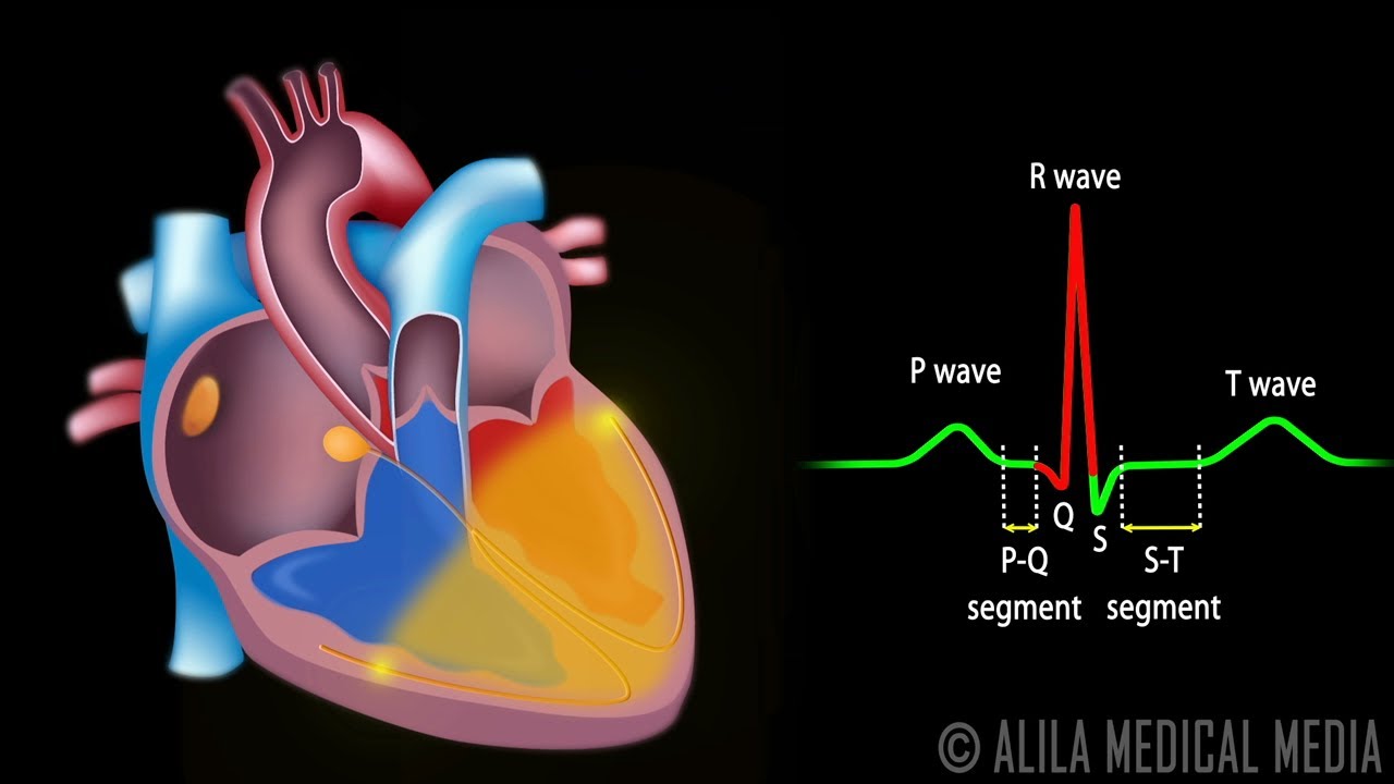
The cardiac conduction system consists of the following components:
The sinoatrial node, or SA node, located in the right atrium near the entrance of the superior vena cava. This is the natural pacemaker of the heart. It initiates all heartbeat and determines heart rate. Electrical impulses from the SA node spread throughout both atria and stimulate them to contract.
The atrioventricular node, or AV node, located on the other side of the right atrium, near the AV valve. The AV node serves as electrical gateway to the ventricles. It delays the passage of electrical impulses to the ventricles. This delay is to ensure that the atria have ejected all the blood into the ventricles before the ventricles contract.
The AV node receives signals from the SA node and passes them onto the atrioventricular bundle AV bundle or bundle of His.
This bundle is then divided into right and left bundle branches which conduct the impulses toward the apex of the heart. The signals are then passed onto Purkinje (purKINjee) fibers, turning upward and spreading throughout the ventricular myocardium.
Electrical activities of the heart can be recorded in the form of electrocardiogram, ECG or EKG. An ECG is a composite recording of all the action potentials produced by the nodes and the cells of the myocardium. Each wave or segment of the ECG corresponds to a certain event of the cardiac electrical cycle.
When the atria are full of blood, the SA node fires, electrical signals spread throughout the atria and cause them to depolarize. This is represented by the P wave on the ECG. Atrial contraction , or atrial systole (SIStoelee) starts about 100 miliseconds after the P wave begins.
The PQ segment represents the time the signals travel from the SA node to the AV node.
The QRS complex marks the firing of the AV node and represents ventricular depolarization:
Q wave corresponds to depolarization of the interventricular septum.
R wave is produced by depolarization of the main mass of the ventricles.
S wave represents the last phase of ventricular depolarization at the base of the heart.
Atrial repolarization also occurs during this time but the signal is obscured by the large QRS complex.
The ST segment reflects the plateau in the myocardial action potential. This is when the ventricles contract and pump blood.
The T wave represents ventricular repolarization immediately before ventricular relaxation, or ventricular diastole (dyASStoelee).
The cycle repeats itself with every heartbeat.
All images/videos by Alila Medical Media are for information purposes ONLY and are NOT intended to replace professional medical advice, diagnosis or treatment. Always seek the advice of a qualified healthcare provider with any questions you may have regarding a medical condition.
Cardiac Axis in 5 min
The basics of the cardiac axis explained in 5 min

กราฟหัวใจ(EKG)มองเส้นยึกๆให้เห็นเป็นรูปได้ยังไง?
ทำตามที่ทุกคนขอมา ยังไม่ต้องลงไประดับการอ่าน แต่แค่ทำความเข้าใจก่อนว่ากราฟบอกอะไรเราได้บ้าง ถ้าอยากให้สอนมากกว่านี้ ขอดูจำนวนlikeก่อน มีเยอะเด๋วสอนต่อ
==============ช่องทางสนับสนับสนุนจ้า==============
►Donate (PayPal/VISA) https://goo.gl/56Jm9Q
►Donate (Truemoney ) https://www.tmtopup.com/topup/?uid=199830
==============ช่องทางติดต่อค้าบ==============
►Facebook Fanpage : https://www.facebook.com/OongStudio/

BASIC EKG Part (1/3) | การแปลผลคลื่นไฟฟ้าหัวใจ
BASIC EKG Part (1/3) | การแปลผลคลื่นไฟฟ้าหัวใจ
การแปลผลคลื่นไฟฟ้าหัวใจแบบพื้นฐานในส่วนของ Part 1
ประกอบด้วย
•กลไกการเกิดคลื่นไฟฟ้าหัวใจ
•การติด EKG 12 leads รวมถึงกรณี 15 leads
•ส่วนประกอบของ EKG
•Normal Sinus Rhythm
•Sinus Bradycardia
•Sinus Tachycardia
•Sinus Arrhythmia
•Sinus Arrest
ติดตาม Part 2 ได้เร็วนี้ค่ะ
ติดตามเพจ
FB : Thebook2hand
IG : Thebook2hand
YouTube : Yingyaichannel

การตรวจคลื่นไฟฟ้าหัวใจ EKG

นอกจากการดูหัวข้อนี้แล้ว คุณยังสามารถเข้าถึงบทวิจารณ์ดีๆ อื่นๆ อีกมากมายได้ที่นี่: ดูวิธีอื่นๆGENERAL NEWS
Articles compiled by CASTU. See more articles in category: GENERAL NEWS